Keratoconjunctivitis sicca (KCS) otherwise known as Dry Eye
Keratoconjunctivitis sicca (KCS) results from lack of aqueous tearing. To understand the “dry eye” syndrome, it is necessary to understand the normal health of the cornea as it relates to the tear film. The cornea is the clear, outer windshield of the eye, but like all living tissues it requires oxygen and nutrients. This is not supplied through blood vessels but through the three-layered tear film. The outer most layer is an oily layer supplied by glands in the eyelids. The middle layer is the liquid (aqueous) layer produced by the lacrimal glands that are located in the upper eyelid and in the third eyelid. This is the layer affected in KCS. The inner most layer of the tear film is in direct contact with the cornea and is a mucous layer produced by glands located in the conjunctival membrane.
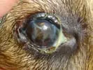
Keratoconjunctivitis sicca allows a breakdown in the corneal tear film and results in patchy, dry areas across the corneal surface or, in more advanced cases, widespread and severe corneal drying. The dried cornea, deprived of oxygen and nutrients through the tear film, rapidly undergoes destructive changes. This drying can lead to corneal ulcers, corneal rupture, ocular infections, significant patient discomfort, corneal scarring, corneal vascularization, and corneal mineralization.
DIAGNOSIS OF KCS
Diagnosis is based on history, clinical signs and a number of diagnostic procedures. These include the Schirmer Tear Testing (STT) which measures watery tear production. Fluorescein stain (a bright green stain) is used to define breaks in the corneal surface and the rate of tear film breakup. Cytology may also be recommended to define the state of health of the conjunctival cells; This has both a prognostic and therapeutic value.
CAUSES OF KCS
A number of causes have been documented for KCS. These include hypothyroidism, infections of the lacrimal glands (ex: canine distemper virus), immune-mediated disease which destroys the tear secreting glands, neurologic conditions and others. Another frequent cause of keratoconjunctivitis sicca is a toxic effect produced by some sulfa-containing antibiotics and non-steroidal anti-inflammatory drugs. Because some of these drugs may be necessary for the treatment of other diseases, it may not be possible to change the patient’s medications and therefore, occasionally the keratoconjunctivitis sicca must be controlled despite being caused by other therapies.
TREATMENT OF KCS
There are several considerations in treating keratoconjunctivitis sicca. A prime consideration is to reduce the overgrowth of bacteria that is common in the dry eye syndrome. The dry eye patient frequently has a buildup of mucous in the eyelids. This mucous is a media for bacterial growth, and antibacterial medications may be warranted. Topical anti-inflammatories are indicated when corneal staining shows no ulceration. This medication helps reduce inflammation of the conjunctival and corneal surfaces and reduces the long term scarring effects. Corticosteroids cannot be used in the face of ulcers because they may decrease healing speed and enhance the ulcerative process. Artificial tear preparations are often indicated to supplement the deficient tear film. In addition to watery preparations, artificial tear ointments are sometimes used to provide prolonged corneal contact overnight and during times that the patient cannot be treated frequently.
New drugs recently developed for the use of treatment of keratoconjunctivitis sicca include immunomodulating agents such as cyclosporine and tacrolimus. These medications are designed to stimulate a patients’ own tear production, and result in the marked increase in tear secretions. The medical treatment for dry eye syndrome generally includes life-long topical medications that are designed to stop progression of the disease and to reverse some of the damage already done. Life-long topical medications seldom give rise to complications, which occur in less than 5% of these cases. Complications of cyclosporine and tacrolimus are typically limited to inflammation of the pink tissue (conjunctivitis) as a result of a local allergic reaction to the medication or the preservatives in the drop. Your awareness of your pet’s symptoms and compliance with recommendations for medication and recheck examinations help control these potential complications.
PROGNOSIS
Most patients with keratoconjunctivitis sicca will do well if medications are administered consistently. In cases where medication cannot be given regularly, surgical techniques such as cyclosporine implantation, parotid duct transposition or even enucleation are considered. Those who select parotid duct transposition need to be aware of potential complications. Any surgical procedure can introduce complications, including potential anesthetic risks. Potential complications include, but are not limited to: break down of the tissue or suture (wound dehiscence), obstruction or deterioration of the salivary gland or duct, salivary stone formation, facial nerve damage, infections at the surgical site, severe periocular wetting, corneal ulcerations, corneal scarring or vascularization. Additionally, surgical procedures that involve the salivary gland, conjunctiva, eyelid or skin surrounding the eye, including parotid duct transposition, can give rise to the following complications: inflammation of the tissue inside the mouth, of the incision on the cheek, of the eyelid (blepharitis), and of the pink tissue (conjunctivitis); corneal mineralization; tearing (epiphora); and secondary moisture-related dermatitis. Some of these complications can lead to blindness.